Back Neck Bones Human - Rear View Of The Bones Of The Torso Stock Photo Alamy / The cervical spine is comprised of the 7 uppermost vertebrae of the vertebral column.
Back Neck Bones Human - Rear View Of The Bones Of The Torso Stock Photo Alamy / The cervical spine is comprised of the 7 uppermost vertebrae of the vertebral column.. Head neck spine back abdomen chest shoulder arm elbow forearm hand wrist lung. Herniated disks or bone spurs in the vertebrae of your neck can press on the nerves branching out from the spinal cord. However, the muscles of the neck can also be easily strained or injured. It runs from the neck to the upper back. Two important reasons why we should know this bone to learn to draw the human back:
Among these structures we can find: Related posts of back of neck bone structure human body bones pics. Human bone is stretching arm and leg (whole body : The motion of the muscles of the neck are divided into four. It runs from the neck to the upper back.
Chronic Neck Pain What You Need To Know from www.drugs.com The occipital bone is the only bone in your head that connects with your cervical spine (neck). Close up macro view of human skull bone showing the anatomy of nasal foramen, nasal septum and orbital cavity. In the back, the neck reaches the c7 vertebra. C3 to c6 are the typical cervical vertebrae characterised by the presence of transverse foramina and, in many people, by their bifid spinous processes. The muscles of the neck provide the mechanism for swallowing, yawning, speaking, and moving the head. Head neck spine back abdomen chest shoulder arm elbow forearm hand wrist lung. An area called the occiput. The motion of the muscles of the neck are divided into four.
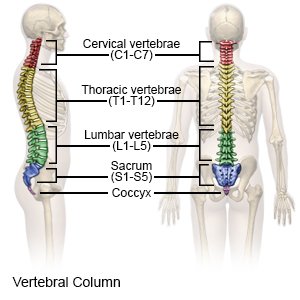
Consisting of 33 bones, more than 120 muscles, over 200 individual ligaments, and an assortment of nerves and supportive discs, the human spine is designed to perform many important functions with maximum efficiency.
Close up macro view of human skull bone showing the anatomy of nasal foramen, nasal septum and orbital cavity. Osteoarthritis causes the cushions (cartilage) between your bones (vertebrae) to deteriorate. Head neck spine back abdomen chest shoulder arm elbow forearm hand wrist lung. Pain in a man's body pain in a man's body on a gray background. A person may refer to the hump on. When you look at the skeleton from behind, you can clearly see the spine running down the back, and the broad plates of the shoulder blades and pelvis. The muscles of the neck run from the base of the skull to the upper back and work together to bend the head and assist in breathing. The cervical spine protects the nerves connecting to. It runs from the neck to the upper back. Neck the neck is the start of the spinal column and spinal cord. The occipital bone is a bone that covers the back of your head; It consists of seven vertebrae. The first cervical vertebra, c1, supports the skull and is named atlas after the greek titan who held the earth on his shoulders.
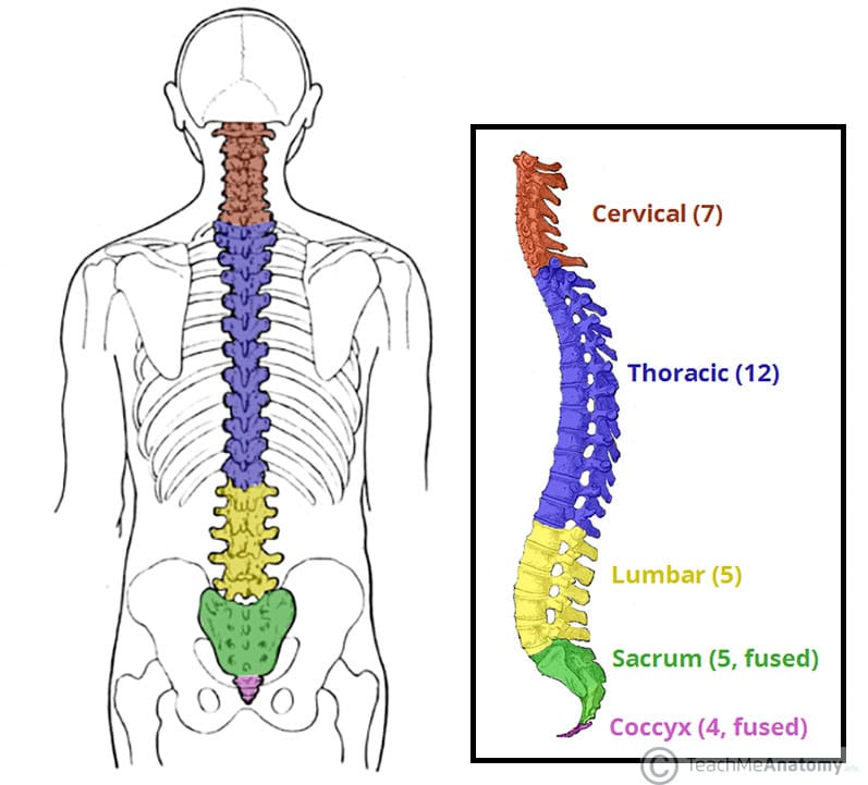
The head rests on the top part of the vertebral column, with the skull joining at c1 (the first cervical vertebra known as the atlas). The human neck is one of the most complex structures we have because it contains many important elements that converge in a very small space. Lateral labeled diagram of the human vertebral spinal column showing vertebrae numbering order and the 5 different regions of the spine. Two important reasons why we should know this bone to learn to draw the human back: The cervical spine and the hyoid bone constitute the bones of the neck.
The Vertebral Column Joints Vertebrae Vertebral Structure from teachmeanatomy.info Researchers have discovered that some young people are developing a bone growth at the back of the skull, due to looking down at their phone. The content of the neck is grouped into 4 neck spaces, called the compartments. Two important reasons why we should know this bone to learn to draw the human back: Neck the neck is the start of the spinal column and spinal cord. The occipital bone surrounds a large opening known as the foramen magnum. The muscles of the neck provide the mechanism for swallowing, yawning, speaking, and moving the head. The human head and neck bones are crucial for structure and support. The skeletal section of the head and neck forms the top part of the axial skeleton and is made up of the skull, hyoid bone, auditory ossicles, and cervical spine.
It runs from the neck to the upper back.
The cervical spine is comprised of the 7 uppermost vertebrae of the vertebral column. The skeletal section of the head and neck forms the top part of the axial skeleton and is made up of the skull, hyoid bone, auditory ossicles, and cervical spine. It runs down the centre of the body. Among these structures we can find: The university of the sunshine. The skull can be further subdivided into: The human head and neck bones are crucial for structure and support. The occipital bone is the only bone in your head that connects with your cervical spine (neck). Herniated disks or bone spurs in the vertebrae of your neck can press on the nerves branching out from the spinal cord. Axial muscles originate on the axial skeleton (the bones in the head, neck, and core of the body), whereas appendicular muscles originate on the bones that make up the body's limbs. The cervical spine is the top part of the spine. Researchers have discovered that some young people are developing a bone growth at the back of the skull, due to looking down at their phone. It anchors muscles of the tongue and throat and holds open the larynx of the respiratory tract.
Among these structures we can find: The axial skeleton is made up of the skull, backbone, breastbone, and ribs. Researchers have discovered that some young people are developing a bone growth at the back of the skull, due to looking down at their phone. The muscles of the anterior neck are arranged to facilitate swallowing and speech. The occipital bone is the only bone in your head that connects with your cervical spine (neck).
Spine Structure Function Parts Segments Spine Problems Spine Health from www.clevelandclinic.org A person may refer to the hump on. The axial skeleton is made up of the skull, backbone, breastbone, and ribs. Browse 3,669 human neck bones stock photos and images available, or start a new search to explore more stock photos and images. The occipital bone is a bone that covers the back of your head; It's easy to take your backbone for granted when everything is working together as expected. The occipital bone surrounds a large opening known as the foramen magnum. And, the clavicle connects to the acromion process of the scapula, creating the acromioclavicular joint. The occipital bone is the only bone in your head that connects with your cervical spine (neck).
Your skeleton can be divided into two main parts.
Contain the common carotid artery, internal. The giraffe's neck is elongated by heterochrony, extension of the time for the embryonic development of these bones. The hyoid is closely associated with the skull but is a floating bone that does not form a joint with any other bone. The skeletal section of the head and neck forms the top part of the axial skeleton and is made up of the skull, hyoid bone, auditory ossicles, and cervical spine. Herniated disks or bone spurs in the vertebrae of your neck can press on the nerves branching out from the spinal cord. The human neck is one of the most complex structures we have because it contains many important elements that converge in a very small space. The occipital bone surrounds a large opening known as the foramen magnum. Related posts of back of neck bone structure human body bones pics. The cervical spine is the top part of the spine. The first cervical vertebra, c1, supports the skull and is named atlas after the greek titan who held the earth on his shoulders. However, the muscles of the neck can also be easily strained or injured. The muscles of the anterior neck are arranged to facilitate swallowing and speech. And, the clavicle connects to the acromion process of the scapula, creating the acromioclavicular joint.


0 Komentar